Showing posts with label Image of the Month. Show all posts

Image of the Month - Pathology

A woman in her early 30s presented to the renal clinic with sudden onset edema. Her urinalysis revealed 4+ protein and 3+ blood. Dysmorphic red cells were noted on microscopy. Apart from her edema, she had no other significant symptoms or physical findings.

Her serum creatinine was 0.8 mg/dl and her albumin was 1.6 g/dl. Her serologies were entirely negative. The protein/creatinine ratio was 6 and 9 on two occasions. The presumptive diagnosis was IgA nephropathy and she proceeded to a renal biopsy.

Low power view of the renal cortex showed mild chronic parenchymal changes that would not be unusual for someone of her age - 2/20 glomeruli were sclerosed and there was focal tubular atrophy and interstitial fibrosis affecting less than 5 percent of the parenchyma
Her glomeruli looked normal with no evidence of inflammation or mesangial expansion.

There was no staining for IgA. There was minimal IgG staining with equal kappa and lamda.

EM showed diffuse effacement of the foot processes consistent with minimal change disease. But, why did she have hematuria?

The answer is revealed by looking closer at the basement membranes. The harmonic mean thickness of the BM was 196nm which is diagnostic for thin basement membrane disease. The hematuria in this case was a red herring - her acute diagnosis was minimal change disease and she simply had not previously been noted to have hematuria.

TBMN is the commonest cause of persistent hematuria, present in about 1% of the population. It has a benign prognosis and is associated with a positive family history. It can sometimes be difficult to distinguish from forms of Alport's syndrome early in the course of the disease. It results from heterozygous mutations in the COL4A3 and COL4A4 genes. Interestingly, the penetrance of hematuria is only 70% so the absence of a positive family history of hematuria does not definitiely suggest a de novo mutation. Episodes of macrosopic hematuria can follow an acute infection and thus mimic IgA although this is relatively rare. One of the bigger issues is making the diagnosis and criteria and measurement techniques vary by institution.

There is no specific treatment required although patients do need to be followed up as there are some atypical cases of Alport's syndrome which can present similarly but are associated with progression. Some have advocated treating patients with ACEi to prevent episodes of hematuria but the evidence for this is not strong. Patients with TBMN can be kidney donors although they should have a renal biopsy first to make certain that there is no other diagnosis. Here is an excellent review from 2006 and more recently, expert guidelines on the management of Alport's and TBMN were published in JASN.

Pathology Case of the Month

An elderly woman presented for evaluation to the rheumatology service with arthritis and neuropathy. She was hypertensive and had microscopic hematuria with dysmorphic red cells on microscopy. Her creatinine was 1.6 which had not changed in the previous year. She had no history of diabetes. Her serology was notable for a positive ANCA, elevated CRP and ANA 1:640. The sense was that despite the presence of the ANCA, the likelihood of a vasculitis was low due to the indolent course but she went on to have a renal biopsy.

A low power view of the renal cortex revealed an obvious nodular appearance of the glomeruli. The tubules were relatively well-preserved.
High-power view of the glomerulus was characteristic of nodular glomerulosclerosis. Notably, there was no evidence of any crescents and no thickening of the capillary loops.
 IF was completely negative

There were no medullary or BM inclusions on EM

Nodular glomerulosclerosis is classically associated with diabetes and this is the first diagnosis that came to the mind of the pathologist when the slides were processed. However, the patient had no history of diabetes. Alternative diagnoses include chronic MPGN and dysproteinemias. There was no evidence of either in this case. The final diagnosis was "Idiopathic Nodular Glomerulosclerosis". This is somewhat of a misnomer as these days, it is thought that IGN is directly related to smoking. The mechanism is uncertain but is suggested in the flow-chart below from an a review in JASN on the topic.


This is not a benign condition. In the largest case series to date of 23 patients with biopsy-proven IGN, 6 patients reached ESRD in a median of 26 months. Predictors of progression included not quitting smoking, lack of ACEi use and the degree of atrophy, fibrosis and arteriosclerosis on renal biopsy.

(Click on any image to enlarge)

Image of the Month - 2

A man in his 50s with a history of diabetes, renal stones, gastric bypass surgery and CKD stage III/IV presented to the clinic with fevers, chills and vague abdominal pain. A urine culture was positive for E Coli and he was treated with a quinolone with resolution of his symptoms. In view of his previous history of renal calculi, a CT abdomen was ordered.


The CT scan revealed a 5 x 2.2 cm mass lesion in the right renal/suprarenal region inseparable from the kidney and the right adrenal gland and the differential diagnosis was either a RCC or adrenal carcinoma. He then proceeded to an MRI abdomen.


This again showed a mass in the right suprarenal region, inseparable from the right kidney and adrenal gland with some central necrosis. At this point, the decision was made to proceed with a partial nephrectomy for likely carcinoma.


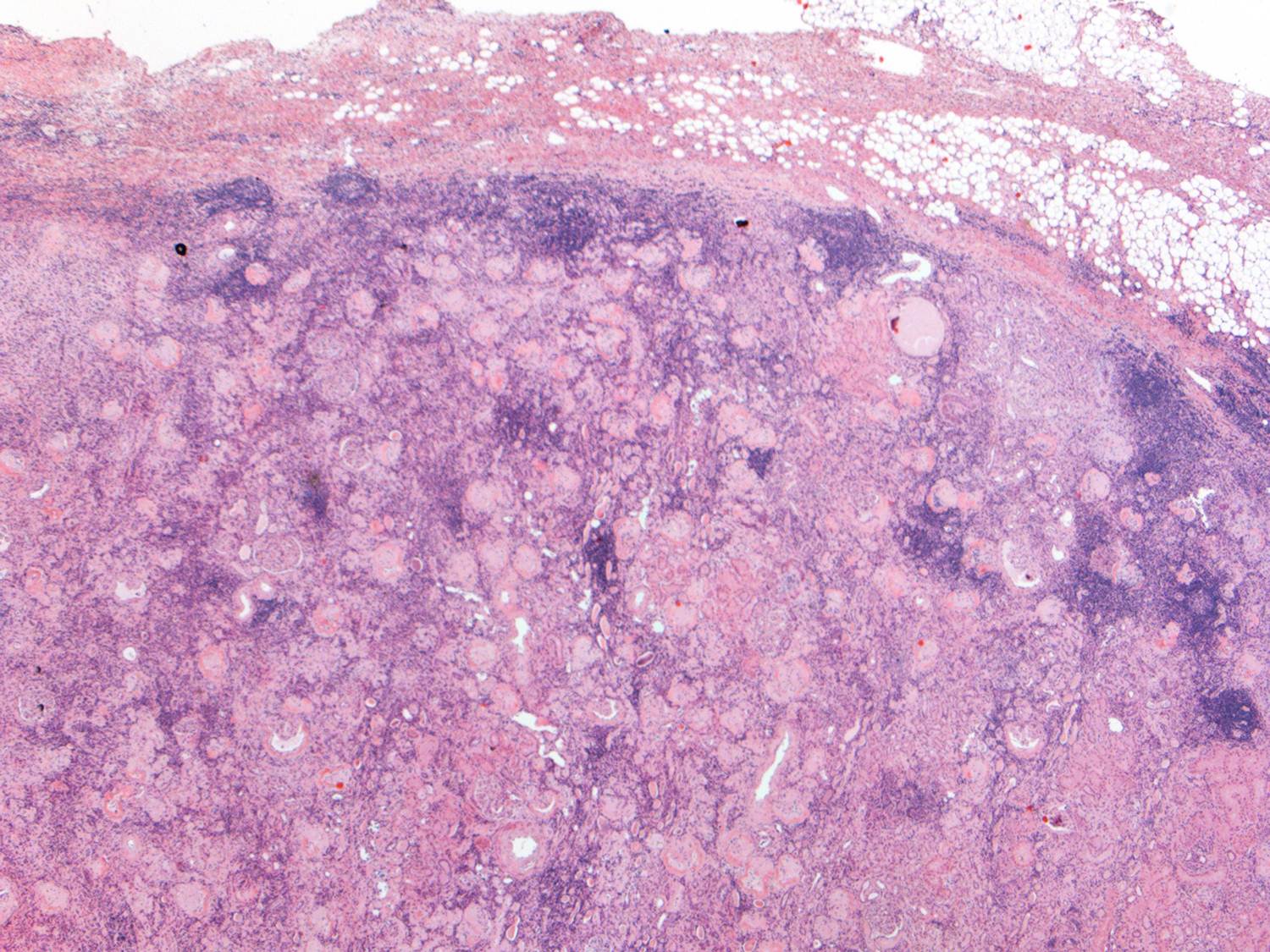
This is a low-power view of the renal cortex. There is diffuse global glomerulosclerosis involving approximately 80% of glomeruli. There is also significant tubular atrophy and interstitial fibrosis with associated areas of inflammation.
There was focal perirenal and intrarenal scarring with disruption of the renal capsule.
The adrenal gland was essentially normal apart from some focal inflammation and adhesion to the capsule. There was no evidence of any malignancy and it is likely that the changes seen were a result of infection which had resolved by the time of the surgery.
Higher power view of the preserved glomeruli revealed changes characteristic of diabetic nephropathy - nodular glomerulosclerosis.
Interestingly, he also had many tubules containing oxalate crystals, likely related to his previous gastric bypass surgery. There was associated acute tubular injury.

This case lead to an interesting debate in our conference. Should he have been treated for a longer period with antibiotics and then rescanned prior to the resection. In the end, the consensus was that the treatment he received was appropriate. Multiple imaging studies were done that were suggestive of malignancy and he had constitutional symptoms including weight loss and fever. A review of all tumor nephrectomies performed at BWH a few years ago revealed that 1/110 cases was not actually tumor. It is hard to argue in that setting that delaying the resection is the appropriate management. Of course, in this case, there was the added complication of advanced CKD and he has now lost some of his residual GFR (although his creatinine has returned to the pre-surgery baseline).

One final image: on the MRI scan, he was incidentally found to have multiple gallstones - I just thought that the picture looked really cool.
Click on any image to enlarge

Image of the Month

The renal services were consulted on a man in his late 70s with a distant history of colon cancer, recent pneumonia and NSTEMI, for investigation of worsening renal function. He had sub-nephrotic range proteinuria (2-3 g/24 hours) and an elevated creatinine (2.3 mg/dl). His serum albumin was 1.8 g/dl. Serologies revealed a negative SPEP and UPEP but a positive p-ANCA (MPO 354). His clinical condition deteriorated and he was transferred to the ICU. Notably, his urine sediment contained muddy-brown and granular casts with no dysmorphic red cells and no red cell casts.

At this point, the differential diagnosis included a vasculitis, drug-induced vasculitis (although this is usually associated with higher ANCA titers) or ATN. We proceeded to a renal biopsy.


Low power view of the biopsy specimen reveals the presence of an obvious medium-sized vessel - likely an arcuate artery

Higher power view of the cortex revealed relatively normal-appearing glomeruli with no inflammation and no crescents. There were some chronic changes with approximately 11% globally sclerosed glomeruli but this was not thought to be related to the current presentation.
There was a dense interstitial infiltrate with many plasma cells and occasional eosinophils. The small arterioles looked normal.

There were two sections of arcuate artery on the specimen:

The first section showed some arteriosclerosis and some perivascular inflammation but no vasculitis


This is a two views of the same section of a medium-sized artery. There is massive infiltration of the vessel wall characteristic of a vasculitis -specifically this has the appearance of polyarteritis nodosa. There was significant fibrin deposition on IF within the walls of the vessel. There was also some mesangial deposition of IgG and IgM.

This is a fascinating case. First, this form of vasculitis is not usually associated with a positive ANCA test and this may have been a red herring. Second, the smaller vessels were normal and if the arcuate artery was not present on the specimen, this patient would likely have been diagnosed with an interestitial nephritis. The proteinuria in this case is probably a result of reduced tubular reabsorption given the fact that there is no significant glomerular disease. The low serum albumin was most likely due to GI losses rather than renal.

Bonus History of Nephrology Point:
Although we associate interstitial nephritis with drug use and know that it was classically described in the setting of methicillin use, AIN was initially described in the setting of acute sepsis. Councilman nephritis was first described in 1898 in autopsy specimens of patients who died with sepsis. Given the plethora of drugs that most septic patients are exposed to these days prior to any biopsy, this is a difficult diagnosis to make at this point but it should be remembered that not all AIN is drugs. The image below is a plate from that paper which is available for free online.


Click on any image to enlarge

Image of the Month - Pathology

A 60 year-old woman with a longstanding history of hypertension, scleroderma and MGUS presented to the emergency room with diarrhea, vomiting and AKI (creatinine increased from baseline of 1.5mg/dl to 3.1mg/dl). She had a distant history of membranous nephropathy diagnosed on a renal biopsy 25 years previously. She had been worked up in the renal clinic for CKD and had a renal US showing relatively small kidneys. Her medications included an ACE inhibitor.

On admission, she had no hematuria. Her BP was elevated although she had a significant postural drop. She had dipstick proteinuria and she was empirically started on oral steroids for a possible GN. A renal biopsy was performed:


The image above is of the renal cortex. A single glomerulus is seen (A) which is hypoperfused. There is significant dilatation of the tubules (B) indicating acute tubular injury. There is attenuation and degeneration of the tubular epithelial cell layer. At the lower end of the image is an atrophic tubule with a hyaline cast.


This image again shows a hypoperfused glomerulus. However, the main finding here is an extremely damaged arteriole (A). There is multilayering within wall of the vessel (that was replicated throughout the biopsy). The vascular lumen is almost occluded with endocapillary proliferation and remodeling of the vessel wall.

In some areas of the biopsy (better perfused), the glomeruli looked relatively normal. There was some mesangial expansion with irregular capillary loops but no evidence of membranous disease.


IF showed some minimal deposition of IgG in the mesangium but equal kappa and lamda light chains.


Finally, the EM showed multiple mesangial deposits (A) of uncertain significance but again, no evidence of membranous disease. Notably, there were was no evidence of active inflammation in the biopsy.

Following the report of the biopsy, the steroids were stopped. The patient's blood pressure was controlled and her renal function returned to baseline within a few days. It is likely that this acute episode was related to volume depletion and acute tubular injury exacerbated by her severe underlying vascular disease. Interestingly, she now has no albuminuria and she is back on her ACEi. The significance of the mesangial deposits remains unclear and she will be followed on an ongoing basis in the renal clinic.

We often see patients like this on consult who present with AKI following a GI illness while on an ACEi. However, we don't normally get to see the pathology in these very common case.

Click on any image to enlarge

Image of the Month - GN with a twist


A man in his 30s with no significant medical history presented to the ED following a fainting episode. Routine screening found an abnormal creatinine, hematuria and proteinuria. Serological work-up including complements were negative. In view of a slowly increasing creatinine and 3g of proteinuria, he was referred for a renal biopsy.


A low power view of the renal cortex revealed diffuse fibrosis and interstitial inflammation. There was evidence of focal sclerosis with occasional sclerosed glomeruli.


A high power view of a typical glomerulus revealed mesangial expansion and proliferation with normal-appearing capillary loops. There were no cellular crescents and some glomeruli had evidence of focal sclerosis.


Examination of the renal vasculature revealed severe arterial sclerosis and moderate arteriolar sclerosis.

Not unexpectedly, the EM found extensive mesangial deposits with no sub-endothelial or subepithelial deposits and a normal appearing basement membrane. The podocyte morphology was also well preserved.


Top on the list of differentials in this patient was IgA nephropathy and as expected, the immunofluorescence revealed extensive mesangial staining for IgA. At this point, many pathology labs would leave it at this and make a straightforward diagnosis of IgA. However, our lab can't just leave it alone and generally also routinely stains for immunoglobulin light chains. Typically in IgA nephropathy, lambda light chains are more prominent than kappa light chains on IF but the difference is not marked.

In this case, there was almost no staining for kappa and marked lambda staining. This suggests that a monoclonal IgA is present. The serum immunofixation was negative and so far, a bone marrow biopsy has not been done.

Monoclonal IgA is rare and has only been reported in case series in the literature. It is often accompanied by a positive SPEP and is thought, at least in some cases, to be related to a clonal expansion of B-cells. About 20% of myeloma is IgA but this is rarely associated with deposition in the kidney (although this may be due to a lack of biopsies). Anecdotally (speaking to my colleagues), this form of IgA nephropathy is relatively aggressive with rapid progression. This has not been formally studied. The question that arises is how to treat this. Should it be treated with rituximab/steroids/velcade? This is relatively toxic treatment and in the absence of an abnormal bone marrow or serum evidence of a monoclonal gammopathy, it is difficult to make this argument. However, one would wonder about the likelihood of recurrence after renal transplantation. 

In this case, the decision has been made to hold off on aggressive treatment for the moment with regular monitoring of the serum. This is a fascinating case and just goes to show how diverse a condition like IgA can be. 

(Click on any image to enlarge)

Pathology Image of the Month

A 47 year old man with ESRD secondary to a chronic TMA presented 3 months following a renal transplant with a rising creatinine (1.44 mg/dl from a baseline of 0.9 mg/dl). His prograf level had ranged from 5-7. His flow XM was positive although no donor-specific antibody was detectable on a single antigen bead. His H&E stain is seen below.


He has a moderate interstitial infiltrate. (A) marks an enlarged, abnormal nucleus with a Cowdry type B inclusion. These inclusions can be seen both in adenovirus infections and in patients with BK nephropathy. (B) marks a lymphocyte in the proximal tubular epithelium – tubulitis. The absence of significant necrosis makes adenovirus less likely and, given the recent history of a renal transplant, the likeliest diagnosis therefore is BK nephropathy.


This image shows immunohistochemical staining for BK (SV-40) in the renal cortex. There is some cytoplasmic staining in surrounding tubules. However, this is non-specific. There is intense staining in the nuclei of infected cells (which are also enlarged relative to the non-infected cells) and this is highly specific for BK-virus infection.


The final image is a EM image of an infected cell. Because BK-viral infection tends to be focal, it is unusual to actually see this appearance on EM. There is the appearance of a crystalline array of viral inclusions and in BK-virus, these tend to the in the range of 40-50 nm in size. In contrast, the inclusions in patients with adenovirus tend to be larger.

(Click any image to enlarge)

Image of the Month

A man in his early 60s was being treated for metastatic adenocarcinoma of the lung for 2 years. Recently, he had  been complaining of increasing hemoptysis and a PET CT was performed which showed progression of his disease. He presented to the ED with general malaise, edema and abdominal bloating. His creatinine had increased from a baseline of 1.0 to 1.5mg/dl and he had >10g urinary protein daily. A renal biopsy was performed. Light microscopy revealed thickened basement membranes with some double-contour formation but no definite "spikes" Immunofluorescence showed dense granular staining for IgG along the capillary loops with some minor staining in the mesangium. The diagnosis was early secondary membranous glomerulopathy most likely related to the progression of his malignancy. An EM image is shown below (Click to enlarge)
Subepithelial deposits typical of membranous nephropathy are easily seen in the image (A). The overlying podocytes are damaged and the foot processes are effaced. The lower part of the image (B) shows an area at the junction of the capillary loop and the mesangium. Here, a deposit is also present but it appears that the overlying glomerular epithelium degenerated and is sloughing into the lumen. This is likely due to complement activation and the formation of membrane attack complexes induced by the presence of the deposits and is thought to be the cause of the severe proteinuria seen in this patient. It is important to note that this is not associated with inflammation because the complexes are out of the reach of inflammatory cells which is not the case where subendothelial deposits predominate. This case was not entirely typical because there were occasional subendothelial deposits noted throughout the glomerulus although they were few and their significance was uncertain. 


Thanks to Dr Bijol for the Image