Showing posts with label renal pathology. Show all posts

Lonely Glomeruli

One of the difficulties in doing molecular research on the kidney is the diversity highly specialized cells that exist in the glomerulus. As a result, it is important to be able to isolate glomerular tissue from the surrounding kidney. A recent paper in KI detailing a method for isolated podocytes reminded me of a relatively simple technique that I was taught a few years ago for glomerular isolation in mice.

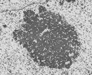
The technique was first described in this paper from 2002 but in brief, it involves injecting the mouse heart with deactivated magnetic beads (after euthanizing them of course). Some of these beads (which are just 5µm in diameter) get trapped in the glomerular capillaries. The kidneys are then removed, minced, digested and passed through a 100µm strainer to remove any larger particulate matter. Finally, the remaining tissue is suspended and exposed to a magnet to pull the glomeruli (with the beads inside) out of the mixture. The glomeruli are then left stuck to the wall of the tube next to the magnet and they can be easily removed.

The picture below is a low-power view of the glomeruli following isolation. You can see that there is very little non-glomerular tissue present, which is remarkable given that the glomeruli make up such a small proportion of renal tissue.


Below is a higher power view of 3 more glomeruli following isolation. You can clearly see the microbeads trapped in the glomerular capillaries. Cool science. (Click on any image to enlarge)

Pathology Case of the Month

An elderly woman presented for evaluation to the rheumatology service with arthritis and neuropathy. She was hypertensive and had microscopic hematuria with dysmorphic red cells on microscopy. Her creatinine was 1.6 which had not changed in the previous year. She had no history of diabetes. Her serology was notable for a positive ANCA, elevated CRP and ANA 1:640. The sense was that despite the presence of the ANCA, the likelihood of a vasculitis was low due to the indolent course but she went on to have a renal biopsy.

A low power view of the renal cortex revealed an obvious nodular appearance of the glomeruli. The tubules were relatively well-preserved.
High-power view of the glomerulus was characteristic of nodular glomerulosclerosis. Notably, there was no evidence of any crescents and no thickening of the capillary loops.
 IF was completely negative

There were no medullary or BM inclusions on EM

Nodular glomerulosclerosis is classically associated with diabetes and this is the first diagnosis that came to the mind of the pathologist when the slides were processed. However, the patient had no history of diabetes. Alternative diagnoses include chronic MPGN and dysproteinemias. There was no evidence of either in this case. The final diagnosis was "Idiopathic Nodular Glomerulosclerosis". This is somewhat of a misnomer as these days, it is thought that IGN is directly related to smoking. The mechanism is uncertain but is suggested in the flow-chart below from an a review in JASN on the topic.


This is not a benign condition. In the largest case series to date of 23 patients with biopsy-proven IGN, 6 patients reached ESRD in a median of 26 months. Predictors of progression included not quitting smoking, lack of ACEi use and the degree of atrophy, fibrosis and arteriosclerosis on renal biopsy.

(Click on any image to enlarge)

Image of the Month - GN with a twist


A man in his 30s with no significant medical history presented to the ED following a fainting episode. Routine screening found an abnormal creatinine, hematuria and proteinuria. Serological work-up including complements were negative. In view of a slowly increasing creatinine and 3g of proteinuria, he was referred for a renal biopsy.


A low power view of the renal cortex revealed diffuse fibrosis and interstitial inflammation. There was evidence of focal sclerosis with occasional sclerosed glomeruli.


A high power view of a typical glomerulus revealed mesangial expansion and proliferation with normal-appearing capillary loops. There were no cellular crescents and some glomeruli had evidence of focal sclerosis.


Examination of the renal vasculature revealed severe arterial sclerosis and moderate arteriolar sclerosis.

Not unexpectedly, the EM found extensive mesangial deposits with no sub-endothelial or subepithelial deposits and a normal appearing basement membrane. The podocyte morphology was also well preserved.


Top on the list of differentials in this patient was IgA nephropathy and as expected, the immunofluorescence revealed extensive mesangial staining for IgA. At this point, many pathology labs would leave it at this and make a straightforward diagnosis of IgA. However, our lab can't just leave it alone and generally also routinely stains for immunoglobulin light chains. Typically in IgA nephropathy, lambda light chains are more prominent than kappa light chains on IF but the difference is not marked.

In this case, there was almost no staining for kappa and marked lambda staining. This suggests that a monoclonal IgA is present. The serum immunofixation was negative and so far, a bone marrow biopsy has not been done.

Monoclonal IgA is rare and has only been reported in case series in the literature. It is often accompanied by a positive SPEP and is thought, at least in some cases, to be related to a clonal expansion of B-cells. About 20% of myeloma is IgA but this is rarely associated with deposition in the kidney (although this may be due to a lack of biopsies). Anecdotally (speaking to my colleagues), this form of IgA nephropathy is relatively aggressive with rapid progression. This has not been formally studied. The question that arises is how to treat this. Should it be treated with rituximab/steroids/velcade? This is relatively toxic treatment and in the absence of an abnormal bone marrow or serum evidence of a monoclonal gammopathy, it is difficult to make this argument. However, one would wonder about the likelihood of recurrence after renal transplantation. 

In this case, the decision has been made to hold off on aggressive treatment for the moment with regular monitoring of the serum. This is a fascinating case and just goes to show how diverse a condition like IgA can be. 

(Click on any image to enlarge)

Pathology Image of the Month

A 47 year old man with ESRD secondary to a chronic TMA presented 3 months following a renal transplant with a rising creatinine (1.44 mg/dl from a baseline of 0.9 mg/dl). His prograf level had ranged from 5-7. His flow XM was positive although no donor-specific antibody was detectable on a single antigen bead. His H&E stain is seen below.


He has a moderate interstitial infiltrate. (A) marks an enlarged, abnormal nucleus with a Cowdry type B inclusion. These inclusions can be seen both in adenovirus infections and in patients with BK nephropathy. (B) marks a lymphocyte in the proximal tubular epithelium – tubulitis. The absence of significant necrosis makes adenovirus less likely and, given the recent history of a renal transplant, the likeliest diagnosis therefore is BK nephropathy.


This image shows immunohistochemical staining for BK (SV-40) in the renal cortex. There is some cytoplasmic staining in surrounding tubules. However, this is non-specific. There is intense staining in the nuclei of infected cells (which are also enlarged relative to the non-infected cells) and this is highly specific for BK-virus infection.


The final image is a EM image of an infected cell. Because BK-viral infection tends to be focal, it is unusual to actually see this appearance on EM. There is the appearance of a crystalline array of viral inclusions and in BK-virus, these tend to the in the range of 40-50 nm in size. In contrast, the inclusions in patients with adenovirus tend to be larger.

(Click any image to enlarge)

Image of the Month

A man in his early 60s was being treated for metastatic adenocarcinoma of the lung for 2 years. Recently, he had  been complaining of increasing hemoptysis and a PET CT was performed which showed progression of his disease. He presented to the ED with general malaise, edema and abdominal bloating. His creatinine had increased from a baseline of 1.0 to 1.5mg/dl and he had >10g urinary protein daily. A renal biopsy was performed. Light microscopy revealed thickened basement membranes with some double-contour formation but no definite "spikes" Immunofluorescence showed dense granular staining for IgG along the capillary loops with some minor staining in the mesangium. The diagnosis was early secondary membranous glomerulopathy most likely related to the progression of his malignancy. An EM image is shown below (Click to enlarge)
Subepithelial deposits typical of membranous nephropathy are easily seen in the image (A). The overlying podocytes are damaged and the foot processes are effaced. The lower part of the image (B) shows an area at the junction of the capillary loop and the mesangium. Here, a deposit is also present but it appears that the overlying glomerular epithelium degenerated and is sloughing into the lumen. This is likely due to complement activation and the formation of membrane attack complexes induced by the presence of the deposits and is thought to be the cause of the severe proteinuria seen in this patient. It is important to note that this is not associated with inflammation because the complexes are out of the reach of inflammatory cells which is not the case where subendothelial deposits predominate. This case was not entirely typical because there were occasional subendothelial deposits noted throughout the glomerulus although they were few and their significance was uncertain. 


Thanks to Dr Bijol for the Image

Still mysterious: the elusive circulating factor for FSGS


Important new findings were recently published in relation to proteinuria and FSGS, which are definitely of interest to our community.

First, the punch line:

There is new evidence for a “circulating factor” in recurrent FSGS in a fascinating case of a re-transplanted kidney (here)

BUT

There is growing evidence that suPAR is a non-specific marker of kidney disease and therefore not likely to be the “circulating factor.”(here)
In fact, it appears that it is non-specifically found in CKD, and correlates with a declining GFR.


Now for some details:

The re-transplanted kidney

A letter to the NEJM editor (here) describes an amazing case of resolution of recurrent FSGS after re-transplantation. 

A 27 year old patient with primary FSGS receiving a kidney from his healthy 24 year old sister developed proteinuria in the nephrotic range (up to 25 g/day!) within 2 days of transplantation, and had no improvement after plasmapheresis and standard immunosuppressive treatment. A renal biopsy confirmed foot process effacement, the first hallmark of recurrent podocyte damage heralding recurrent FSGS. Incredibly, with all appropriate consents and institutional approval, the transplant team removed the allograft from Patient 1 and re-transplanted it into another patient who had ESRD due to diabetes. Within 3-4 days, the proteinuria resolved and a repeat biopsy showed resolution of foot process effacement and re-establishment of a normal podocyte architecture. Eight months later, Patient 2 is reported to be doing very well, with good allograft function and no proteinuria.

This case demonstrates in a remarkable way that recurrent FSGS results from an elusive “factor” rapidly produced by the recipient (with primary FSGS), and that the allograft itself can remain fully functional if removed from the influence of this “factor” and placed in another patient.

suPAR is not suPER specific

What may have seemed to be exciting news in 2011, namely the notion that soluble uPAR may be predictive of recurrent FSGS (here), appears to be unfortunately evolving into yet another unsuccessful attempt to identify the ever elusive circulating factor.

Recent work published in Kidney International by Maas et al. (here) confirms that suPAR is not able to distinguish between idiopathic FSGS, secondary FSGS or minimal change disease. 

This is actually not surprising, because a closer look at the clinical data in Wei et al. (here) reveals that the admittedly arbitrary cut-off for separating primary FSGS from all other glomerular disease (3000 pg/ml) did not hold up when tested among their patient cohorts with idiopathic, recurrent versus non-recurrent FSGS (all had suPAR> 3000 pg/ml, thus suPAR could not predict the recurrent from the non-recurrent cases). 

The second figure in the Maas et al. paper may help explain this conundrum: they show a negative correlation between suPAR and eGFR, meaning that as GFR drops, suPAR levels rise, which essentially means that suPAR is simply a marker of CKD.

Future work will no doubt continue to address these issues, but the apparent lack of specificity of suPAR for FSGS casts serious doubt on its proposed role as the circulating factor.

So, the search is still on!!!